In the field of criminalistics, the analysis of bite marks has evolved from simple photographic comparison to a high-precision metrological process. An injury left by a bite is no longer documented with rulers and film; today, intraoral scanners and photogrammetry allow capturing the topography of the lesion and the suspect's dentition in a three-dimensional digital environment, eliminating perspective bias and soft tissue distortion.
Technical Workflow: Capture, Alignment, and Overlay 🛠️
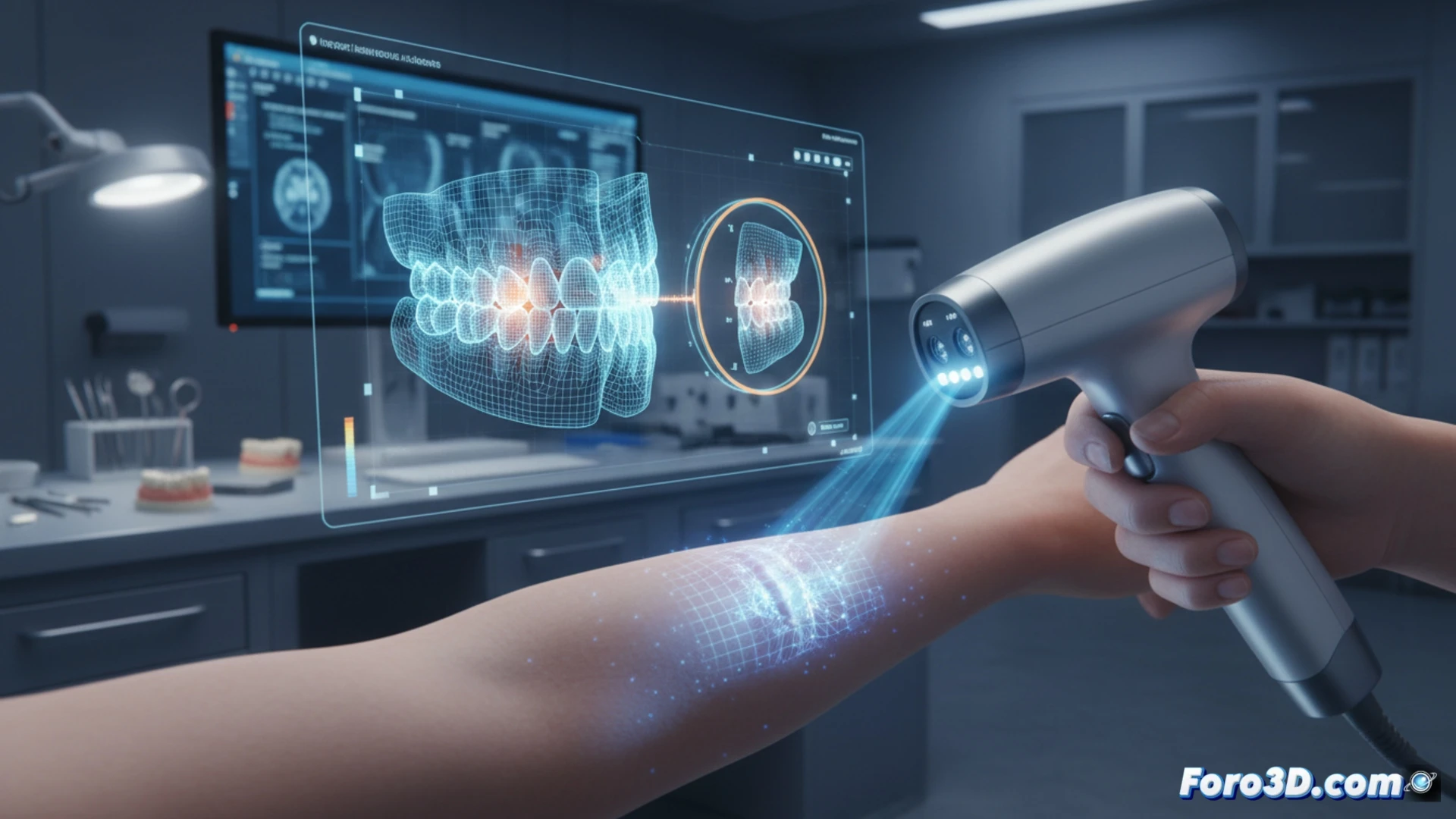
The forensic protocol begins with capturing the bite mark on the victim's skin using high-resolution photogrammetry, generating a textured polygonal mesh that reflects tissue deformation. Simultaneously, an intraoral scan of the suspect is performed using devices such as the 3Shape TRIOS or Medit i700, obtaining a digital model of the dental arches. Software like 3D Slicer or CloudCompare allows aligning both geometries using ICP (Iterative Closest Point) algorithms. The final overlay reveals incisal edge matches, dental rotation, and wear patterns that are quantifiable in microns, generating an expert report that includes chromatic deviation maps.
The Challenge of Living Tissue and Expert Reliability 🔬
Despite technical precision, the greatest challenge remains the elasticity of human tissue. The skin deforms and recovers, forcing experts to work with biomechanical simulation models to estimate the original position of the bite. Real cases, such as the 2022 Florida assault trial, demonstrated that 3D scanning reduced the error margin from 40% to less than 5% in identification. The technology does not replace the expert, but provides them with a statistical validation tool that withstands judicial scrutiny.
Since 3D scanning of bite marks eliminates the subjectivity of visual comparison, what specific challenges does it present in the digital chain of custody to be admissible as evidence in a trial?
(PS: don't forget to calibrate the laser scanner before documenting the scene... or you might be modeling a ghost)