The recent news about the failure of a vertebral hydrogel has put the 3D biomedicine community on alert. This biocompatible material, designed to replace damaged discs, showed premature cracks under load. To understand the origin of the collapse, engineers have turned to three-dimensional modeling technologies that allow them to digitally replicate the biomechanics of the implant and the affected vertebra.
Digital twin and stress simulation 🧬
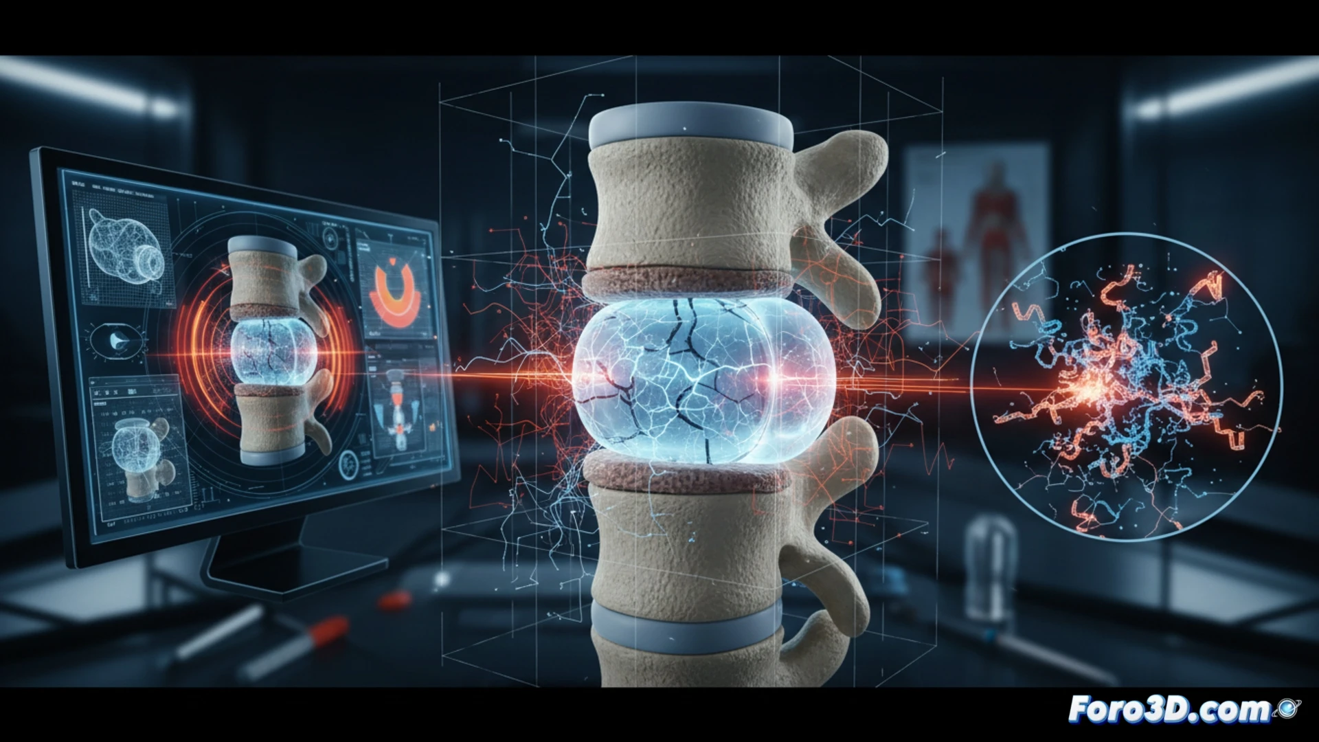
The process begins with a micro-CT scan of the vertebra and the failed hydrogel. With this data, an exact digital twin is generated that reproduces the geometry of the implant and its porous microstructure. Using finite element software, the typical axial and torsional loads of the lumbar spine are applied. The simulation reveals that the failure originated in areas of high stress concentration, where the hydrogel crosslinking was insufficient. This virtual analysis avoids destructive testing and accelerates the diagnosis of mechanical failure.
Surgical redesign assisted by 3D printing 🛠️
With the simulation data, surgeons modify the internal architecture of the hydrogel, adding reinforcement channels and varying the density of the polymer mesh. A 3D prototype is printed with a test material to verify the fit with the patient's vertebra. This physical model allows planning the revision surgery with millimeter precision, reducing the risk of a new failure and optimizing the integration of the implant into the spine.
What role does finite element simulation play in predicting critical points of mechanical fatigue within 3D printed vertebral hydrogels before their in vivo implantation
(PS: and if the printed organ doesn't beat, you can always add a little motor... just kidding!)