When Your 3D Models Could Actually Beat
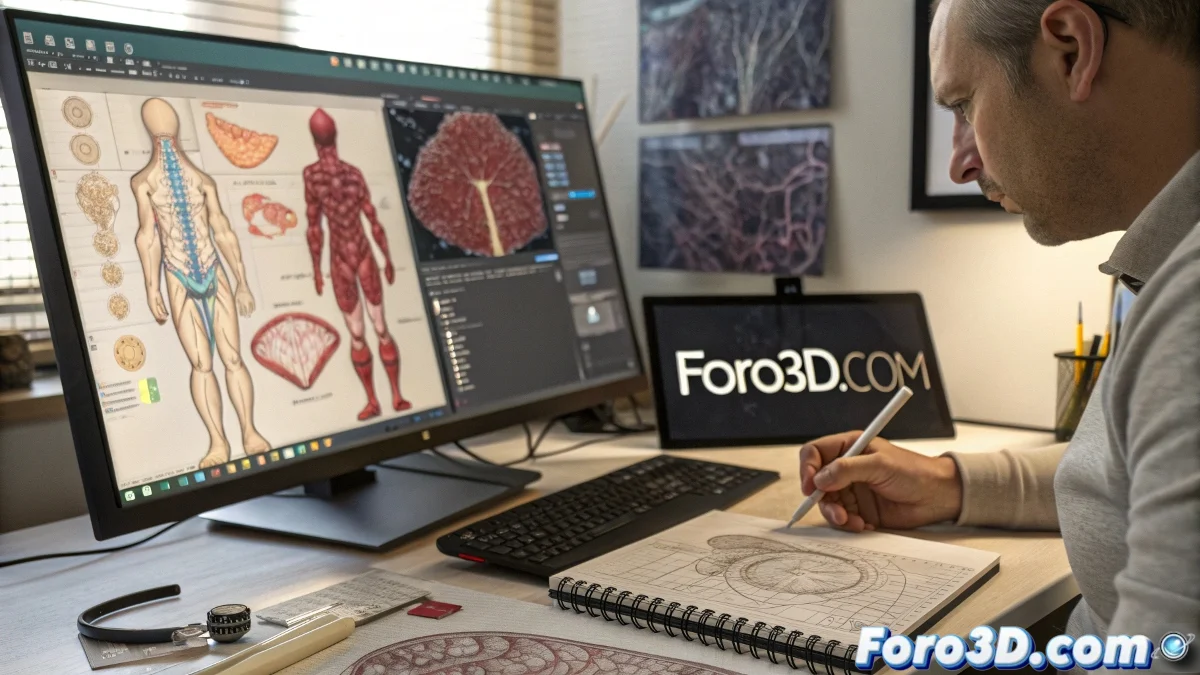
Carnegie Mellon's vascular bioprinting isn't just a medical breakthrough - it's the point where digital art and life science meet. Those blood vessels we model in Blender could soon have their real-life version. ❤️🖨️
Why This Matters to 3D Artists
- New Applications: Your skills can contribute to medical advances
- Anatomical Accuracy: Modeling with precision was never so crucial
- Job Opportunities: Medical visualization is a growing field
- Technical Inspiration: Extreme organic modeling challenges
"I modeled a circulatory system for a game and now it turns out it could help save lives... does this count as medical experience on my CV?" - 3D Artist reconsidering their career.
Software for Vascular Modeling
| Task | Tool | Difficulty |
|---|---|---|
| Organic Modeling | ZBrush/Blender | High |
| Flow Simulation | Houdini | Advanced |
| Realistic Texturing | Substance 3D | Medium |
| Interactive Visualization | Unreal Engine | Medium-High |
How to Get Started in 3D Medical Visualization
- Study real vascular anatomy (books, digital atlases)
- Practice modeling complex tubular structures
- Learn basic principles of fluid dynamics
- Explore open medical datasets for reference
- Collaborate with bioprinting communities
The Future is Already Here
Next steps in this convergence:
- Custom 3D models for each patient
- Tissue printing directly from DCC software
- Surgical simulations in virtual reality
- Educational video games with real anatomy
While we wait for Blender to include a "Surgeon Mode", we can start applying our 3D knowledge to projects that truly make a difference. Because in the end, few satisfactions surpass knowing that your models could one day... literally pump life. 💓
And if your 3D printer can't handle collagen yet, at least your renders can show what that future will look like. That said, better not try to print a heart on your Ender 3... for now. 🚑