OsiriX: Visualize and Navigate Medical Images in 3D
OsiriX is a specialized program for processing medical images. Its main function is to load and handle computed tomography (CT) and magnetic resonance imaging (MRI) studies to generate a three-dimensional volumetric representation of the human body. 🏥
Synchronized Multiplanar Reformatting
The central tool of the software is the 3D MPR viewer. When importing a series of DICOM format images, OsiriX builds a volumetric model. The user can move the cutting planes in any of the three main orthogonal views, and the other two update instantly, maintaining perfect spatial correlation. This synchronization helps locate anatomical structures and possible anomalies more quickly.
Main views it generates:- Axial slices: Transverse view from feet to head.
- Coronal slices: Frontal view, dividing the body into anterior and posterior.
- Sagittal slices: Lateral view, dividing the body into left and right.
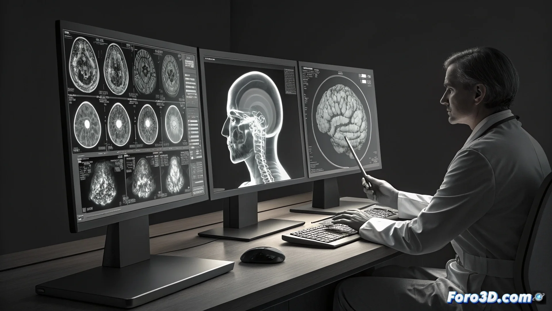
The software displays the data, but the key remains in who analyzes it.
Interface and Tools for Effective Analysis
OsiriX's interface logically organizes the windows of different projections to optimize workflow. It includes a complete set of utilities designed for professionals.
Key features included:- Tools to measure distances, angles, and volumes directly on the images.
- Options to annotate findings and adjust the window level (window/level) to enhance contrast.
- Support for applying various color palettes (look-up tables) and industry-standard formats.
Integration and Target Users
This software integrates with PACS systems (Picture Archiving and Communication System), facilitating the handling of large volumes of studies. It is primarily aimed at radiotherapists and medical researchers who need to analyze imaging data in an advanced way. Its power for diagnosis is a valuable tool, but it does not replace the expertise and judgment of the specialist who must interpret what they visualize. 🔍