OsiriX: Specialized Software for DICOM Medical Image Visualization
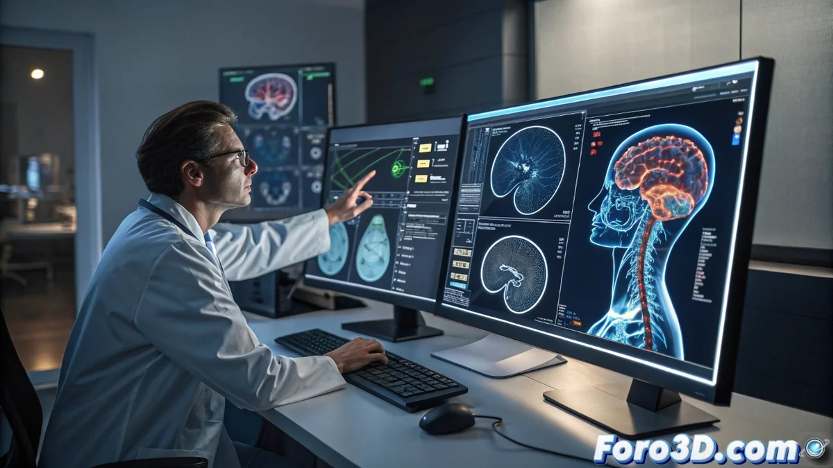
OsiriX represents an open-source solution specifically developed for the visualization and processing of DICOM medical images. This platform has established itself as a fundamental tool in clinical and research environments, facilitating the detailed analysis of computed tomography studies, magnetic resonance imaging, and various diagnostic imaging modalities 🏥.
Advanced Capabilities in Medical Imaging Diagnosis
The software integrates a comprehensive set of tools designed to meet the demands of modern medical diagnosis. Professionals can perform smooth navigation between three-dimensional slices and generate volumetric reconstructions with extraordinary diagnostic precision, allowing for a more comprehensive interpretation of medical studies.
Highlighted Features in Medical Environments:- Precise anatomical measurements and advanced segmentation of body tissues
- Generation of 3D reconstructions from complete series of DICOM images
- Fusion of different imaging modalities to correlate metabolic and anatomical information
OsiriX's ability to transform thousands of tomography slices into interactive three-dimensional models has revolutionized the way specialists approach medical imaging diagnosis.
Fields of Application and User Profiles
This platform is specifically aimed at radiology professionals, surgeons, oncologists, and biomedical researchers who require advanced processing of medical images. Its implementation spans from hospitals for routine clinical diagnosis to research laboratories dedicated to the development of new image analysis algorithms.
Available Versions and Certifications:- OsiriX MD: Certified version for clinical use with full medical validation
- OsiriX Standard: Oriented toward research, medical education, and academic development
- Both versions maintain the core functionalities of 3D visualization and DICOM processing
Impact on Contemporary Medical Practice
OsiriX has radically transformed the experience of reviewing imaging studies, making the analysis of thousands of tomography slices as meticulous as it is intuitive and efficient. The ability to rotate structures in 3D and adjust visualization parameters in real time provides specialists with tools that significantly improve diagnostic precision and intervention planning 🩺.