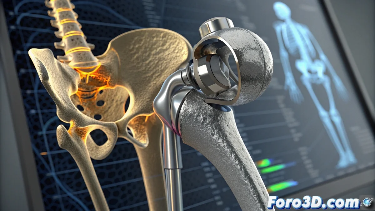
Forensic Analysis of a Hip Implant Using Micro-CT
The path to technical truth begins with a premature failure of a hip prosthesis, an event that plunges the patient into pain and severely limits their mobility. To uncover the underlying cause, the extracted component becomes the key piece in a forensic engineering investigation. The crucial first step is a non-destructive scan using high-end micro-computed tomography systems, such as Nikon CT or Zeiss Metrotom models. This technology generates a high-resolution three-dimensional volumetric representation capable of revealing critical details invisible to the human eye: micro-cracks, material porosity, and wear patterns at the micrometric scale. 🔍
Volumetric Reconstruction and Comparison with the Ideal Design
The raw scanner data is transferred to specialized software like Volume Graphics VGSTUDIO MAX. Here, the volumetric point cloud is processed to isolate the object of interest, remove artifacts, and perform high-precision measurements, including a quantitative porosity analysis. Subsequently, in a platform like Geomagic Control X, this 3D model reconstructed from physical reality is digitally aligned with the original CAD design of the prosthesis. This deviation comparison is fundamental, as it can highlight anomalous wear, permanent deformations, or, conclusively, a discrepancy between what was manufactured and what was designed, pointing directly to a manufacturing defect.
Key Stages of the Reverse Engineering Process:- Data Acquisition: Non-destructive scanning with micro-CT to obtain a high-fidelity volumetric model.
- Processing and Cleaning: Isolation of the implant and removal of noise or artifacts in volumetric analysis software.
- Geometric Comparison: Overlay and deviation analysis between the scanned model and the theoretical CAD plan.
This forensic pipeline converts complex volumetric data into irrefutable technical evidence for a legal process.
Computational Simulation to Validate the Hypothesis
To consolidate the evidence found, the precise digital model can undergo a finite element analysis (FEA) in environments like Abaqus. In this phase, the cyclic loads and real biomechanical conditions to which the joint was subjected are replicated. The fatigue simulation identifies stress concentration zones and predicts the component's lifespan under those conditions. If these calculated high-stress areas spatially match the micro-cracks detected in the micro-CT scan, and the simulation further indicates a premature failure under normal service loads, a solid causal link is established between the defect (whether geometric or material) and the injury suffered by the patient.
Specialized Software Used in the Analysis:- VGSTUDIO MAX (Volume Graphics): For processing, visualization, and quantitative analysis of micro-CT volumetric data.
- Geomagic Control X (3D Systems): For 3D metrology, alignment, and deviation comparison against the reference CAD.
- Abaqus (Dassault Systèmes): For finite element simulation and component fatigue analysis.
From Data to Legal Decision
This comprehensive forensic workflow transforms a failed medical component into a set of objective digital evidence. The combination of non-destructive internal inspection, metrological comparison, and simulation-based validation builds a robust technical case. The next time a suspicious creak is heard in an artificial joint, it could be the start not only of physical discomfort, but of a forensic engineering case destined to determine responsibilities and, ultimately, improve manufacturing and safety standards. ⚖️🦴