3D Computed Microtomography: Key to Differentiating Fractures in Calcined Remains
In the scene of a fire, bone remains present a critical forensic enigma: were the fractures caused by trauma before death or are they simply the result of exposure to extreme heat? 🔥 Resolving this dilemma is essential to establish the cause of death. Fortunately, 3D imaging technology, particularly computed microtomography (Micro-CT), has emerged as a revolutionary tool, offering an unprecedented window into the bone's internal structure with millimeter resolution and in a non-destructive manner.
Ultra-High-Resolution Volumetric Data Acquisition
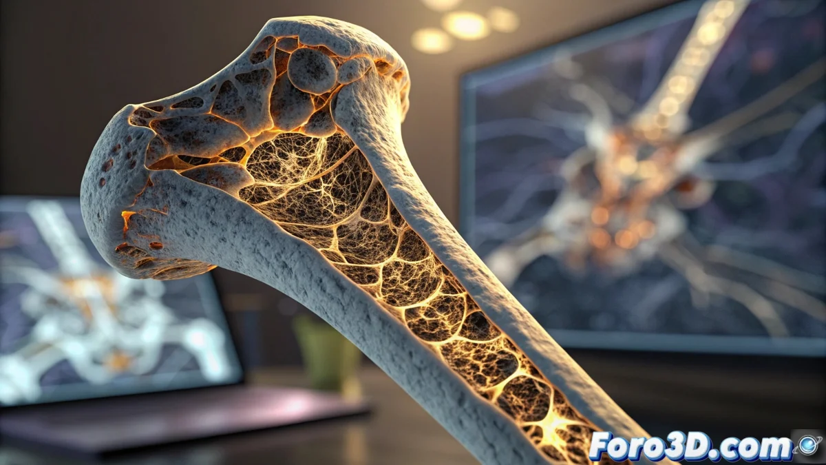
The technical pipeline begins with the digital capture of the sample. Using specialized scanners like Bruker SkyScan systems, thousands of 2D X-ray images are obtained, which, when assembled, generate a high-fidelity three-dimensional volumetric model. This process captures minute details, such as the architecture of the bone trabeculae and microfractures that completely escape visual or conventional radiological inspection. The integrity of the sample is preserved intact, which is vital for subsequent analyses or as evidence.
Key Features of Micro-CT Acquisition:- Micrometric Resolution: Capable of revealing finer internal details than a human hair, essential for identifying the origin of injuries.
- Non-Destructive Method: Preserves the original physical evidence for future reviews or contradictory expert opinions, a fundamental principle in the chain of custody.
- Complete Volumetric Representation: Generates an interactive 3D model that can be virtually sectioned in any plane without damaging the real specimen.
While the fire tries to erase the evidence, 3D technology gives voice to the bones, revealing a story that the heat could not completely consume.
Forensic Processing and Analysis in Specialized 3D Environments
Once the data volume is acquired, the analysis is transferred to scientific visualization software such as Dragonfly, Avizo, or even open-source packages. These platforms allow the forensic anthropologist to perform a comprehensive virtual inspection. The ability to segment, measure, and visualize in augmented reality is what enables the crucial forensic distinction.
Distinctive Patterns Identifiable in 3D:- Perimortem Fractures (Traumatic): Typically present sharp edges, defined angles, and a radial propagation pattern suggesting mechanical impact.
- Thermal Fractures (Fire Product): Exhibit more irregular and curvilinear morphologies, accompanied by signs of shrinkage, carbonization, and a characteristic crazing pattern caused by dehydration and contraction of bone collagen.
- Microstructure Analysis: The study of the trabecular network can show differences in its integrity, providing clues about prior bone health and the direction of applied forces.
The Digital Verdict: Bringing Objectivity to the Investigation
The implementation of this 3D digital workflow transforms forensic anthropology. No longer is reliance solely on the subjective interpretation of surface marks. Instead, there is objective and quantifiable evidence in the form of 3D models, precise measurements, and comparative visualizations. This not only helps to clarify the cause of death, but also strengthens expert testimonies in court, offering a clear and understandable representation of complex findings. The synergy between forensic medicine and 3D visualization technology marks a before and after in the investigation of crimes where fire was used to hide the truth. 🦴