A Biomechanical Model Differentiates Injuries from Falls or Shaking
Distinguishing whether a baby's brain injuries result from an accident or a violent act represents a key forensic challenge. To address it, experts now implement a workflow based on 3D simulation that generates objective digital evidence. This method transforms medical data into dynamic models that reveal the mechanics of the trauma. 🧠
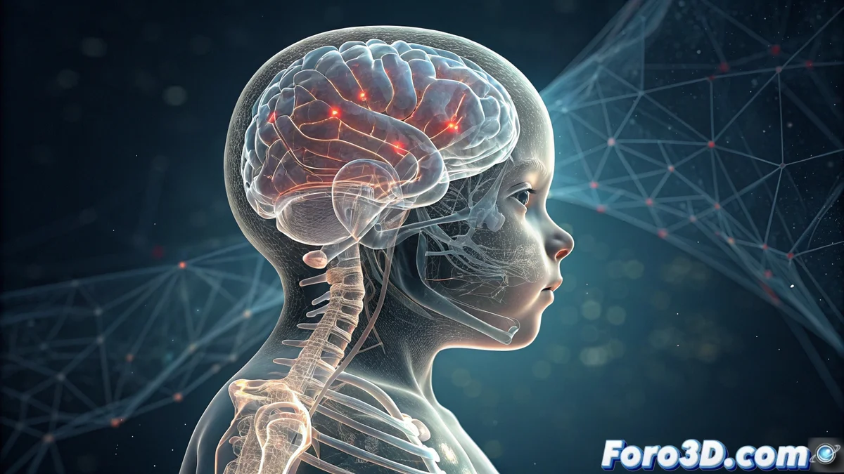
From MRI to Virtual Model
The process begins with the patient's magnetic resonance imaging (MRI) scans. Using specialized software like 3D Slicer, technicians segment and reconstruct the main anatomical structures to create an accurate three-dimensional digital model. This model includes the brain, skull bones, and cervical spine, forming the geometric basis for virtual tests.
Key Phases of Reconstruction:- Import and process the patient's MRI data.
- Segment tissues to isolate the brain, skull, and vertebrae.
- Generate a detailed 3D mesh ready for simulation.
The truth is not always in what is said, but in how the meninges move within a digital file.
Simulating Two Critical Scenarios
The anatomical model is exported to a biomechanical simulation environment like Madymo or LS-DYNA. There, engineers define the properties of biological materials and boundary conditions. Then, they run two separate scenarios: one replicates a fall from a specific height and the other simulates the acceleration and deceleration forces characteristic of violent shaking. The software calculates the shear forces and accelerations impacting the brain tissue in each case.
What the Simulation Calculates:- Inertia and shear forces in the brain parenchyma.
- Stress and strain patterns in the structures.
- Dynamics of cerebrospinal fluid.
Contrasting Virtual Data with Real Evidence
The final simulation result, showing damage and mechanical stress maps, is rigorously compared with the injuries documented in the baby's autopsy or neuroimaging. This objective comparison allows supporting or refuting a hypothesis about the origin of the trauma. Adapting advanced platforms, such as Simulia Living Heart, to model brain dynamics marks an advance in forensic engineering, providing measurable data to a debate that previously relied more on subjective testimonies. 🔍