3D Technology Revolutionizes Animal Bite Forensic Investigation
Methods for analyzing animal attacks are changing radically with the arrival of three-dimensional technology. 🐕🦺 Instead of relying solely on two-dimensional records, experts now capture physical evidence with unprecedented precision, generating objective digital evidence that is crucial in legal processes.
Creating a Precise Digital Record of the Wound
When an attack occurs, the first step is to document the injury with a portable 3D scanner, such as the Artec Eva. This device records the bite and generates an exact three-dimensional model. This model preserves vital metrics: the separation between perforations, how deep they are, and the inclination of each dental mark. Thus, a unique digital fingerprint is formed that serves as fundamental expert evidence.
Key data captured by 3D scanning:- The exact distance between the fang penetration points.
- The depth and contour of each mark in the tissue.
- The angulation and individual characteristics of the dental impressions.
The fidelity of the 3D model turns a wound into objective and quantifiable physical evidence.
Matching the Evidence with the Suspect
The process continues by comparing the digital fingerprint with the possible aggressor. The animal's dentition, for example a dog's, is digitized to obtain a reference 3D model. Using programs like CloudCompare, experts overlay and align both models in virtual space. The goal is to find matches between the wound and the animal's dentition, analyzing shape, size, and tooth arrangement. This method allows confirming or excluding an animal's involvement on a scientific basis.
Software used for comparison:- CloudCompare: For aligning and measuring correspondence between point clouds.
- Geomagic: For processing and analyzing 3D surface overlays with high precision.
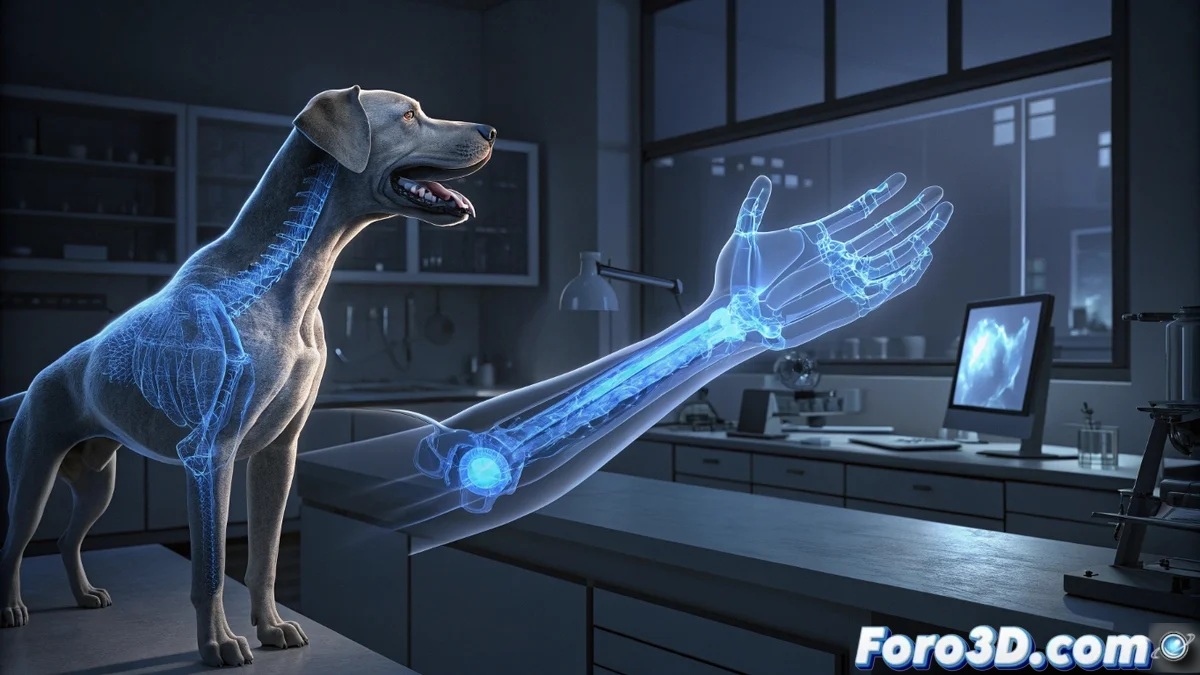
Integrating and Visualizing Complex Medical Information
In intricate injuries, data from the surface scanner is fused with medical images, such as CT scans. Tools like 3D Slicer allow visualizing and segmenting damaged anatomical structures. 🩺 With this software, forensics experts can isolate affected tissue, measure the internal trajectory of the bite, and understand its impact in the complete context of the victim's body. A more robust case is built by combining clinical evidence with the external 3D model.
In short, this methodology offers a compelling forensic conclusion: sometimes, the match between the teeth and the wound is more accurate than any explanation the animal's owner might attempt. 3D technology not only documents but proves with data.