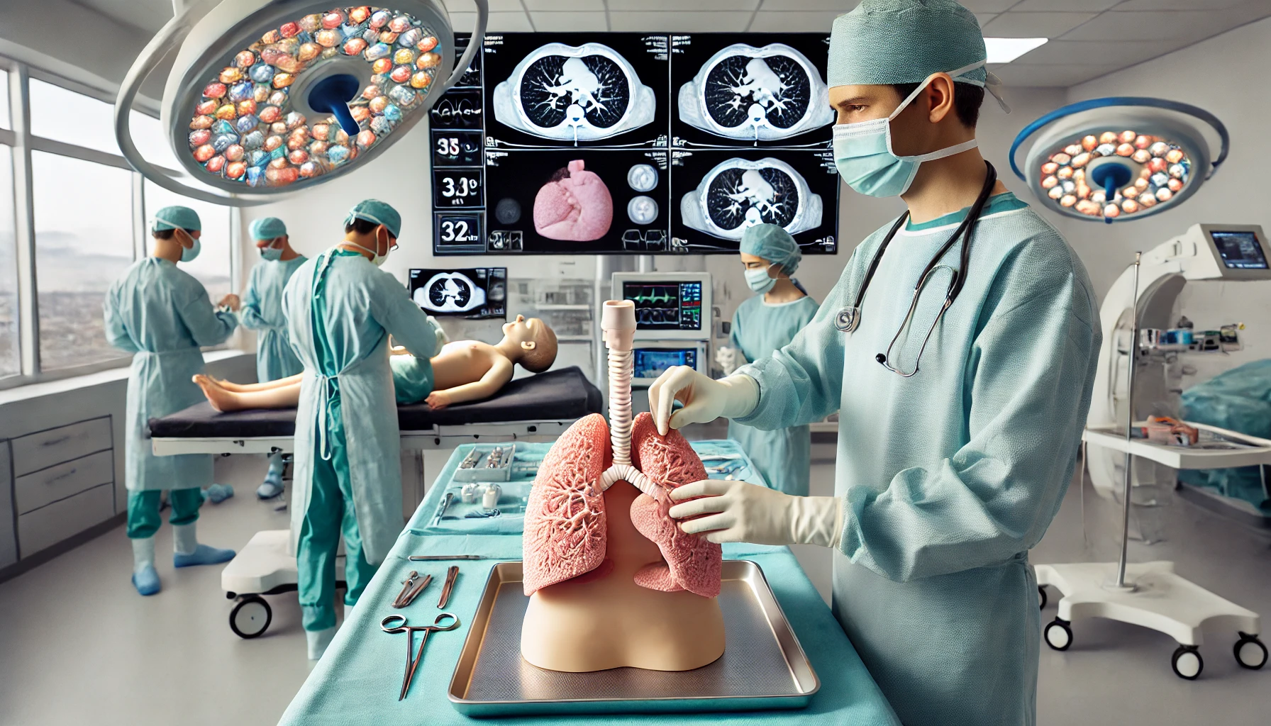
In the field of medicine, preparation prior to a surgical intervention is fundamental to ensure the safety and success of procedures. A hospital in Hong Kong has taken a step forward in this direction by developing 3D printed models of newborns' lungs. These models allow doctors to practice the treatment of pneumothorax, a condition that can cause lung collapse and compromise breathing and cardiac function.
Fusion of Artificial Intelligence and Clinical Data
For the creation of these 3D models, the hospital's research team has combined artificial intelligence with real patient data, including CT scans. To achieve a more realistic texture and properties, the development has also integrated biological data extracted from pork and chicken tissues.
“The combination of 3D printing and artificial intelligence is revolutionizing surgical preparation, improving safety and precision in medical interventions.”
Overcoming Limitations in Medical Training
One of the main challenges in medical training is the scarcity of real cases of pneumothorax in newborns, which makes it difficult for doctors to gain practical experience in its treatment. Thanks to these 3D printed simulations, medical staff can:
- Rehearse the procedure in a controlled environment before applying it to real patients.
- Improve precision in the insertion of chest drains.
- Reduce the risk of complications during surgery.
Precise Training for Delicate Surgeries
The procedure is extremely complex and must be followed with precision. We cannot put the patient at risk due to lack of experience or errors in the steps. It is essential that doctors receive comprehensive training before performing the intervention on a real patient, explains Dr. Víctor Chan, consultant in pediatrics and adolescent medicine at the hospital.
Impact on Patient Safety
The speed in applying the treatment is especially critical in newborns, whose health conditions can deteriorate in minutes without proper intervention. These medical training models provide healthcare professionals with the opportunity to:
- Rehearse each step of the procedure in a safe environment.
- Reduce the margin of error in critical situations.
- Improve the success rate in the treatment of pneumothorax.
A Key Advance in Pediatric Surgery
The use of 3D models in medical training represents a significant advance in pediatric surgery. This technology not only improves the precision and safety of procedures but also opens the door to future applications in the treatment of other critical conditions. With the continuous development of simulation tools, medicine continues to advance toward a future with safer and more effective surgeries.