3D Electron Microscopy Identifies Pollen in Crime Scenes
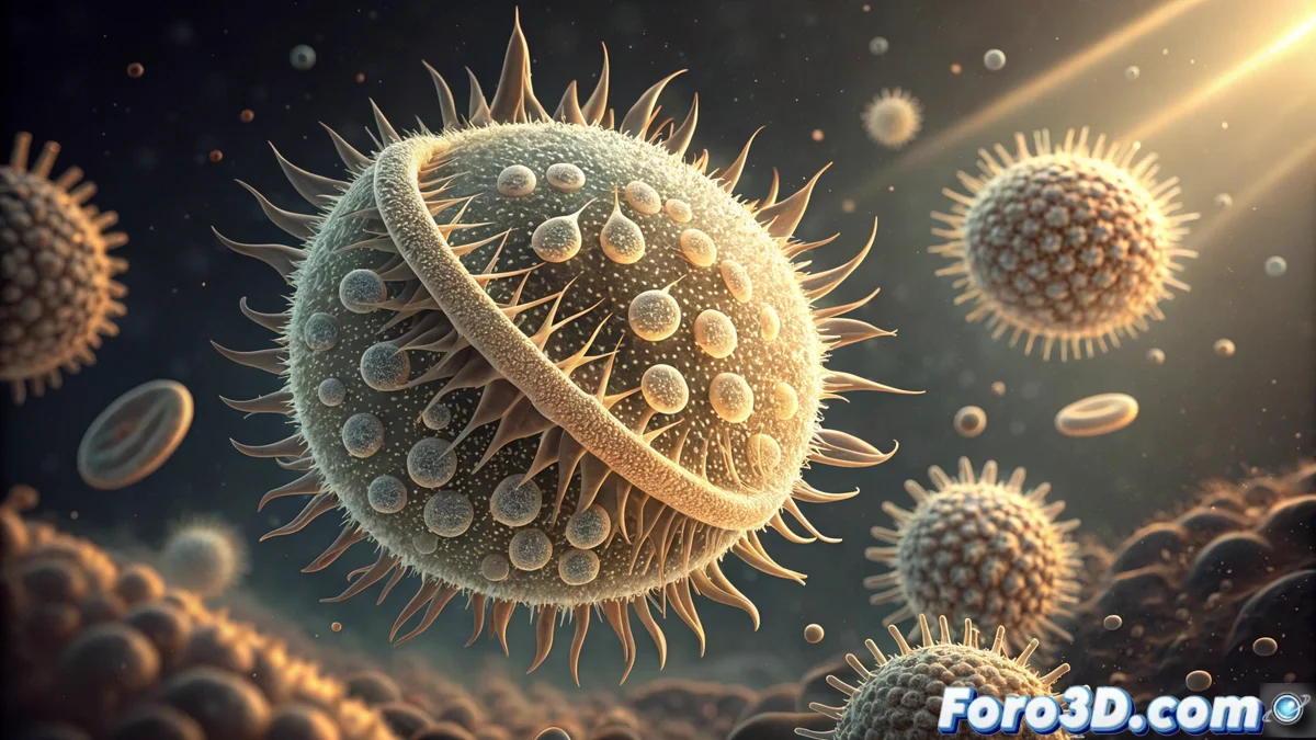
In an investigation, the tiniest evidence is often the key 🔑. A pollen grain found on clothing can point to a specific location. To confirm this link, experts no longer just look at flat images. Now, they use a Scanning Electron Microscope (SEM) to capture the full shape of the particle. This equipment produces hundreds of high-resolution images that, when combined, reveal the three-dimensional structure of the pollen with unprecedented detail. This 3D model becomes a much more powerful comparative proof than traditional two-dimensional photographs.
Technical Process to Reconstruct the Grain in 3D
The workflow starts with the software that controls the SEM, such as those from Zeiss or Thermo Fisher brands, to obtain hundreds of digital slices of the pollen grain. Then, these serial images are imported into specialized 3D reconstruction applications, like Avizo or Dragonfly. These programs process the image stack and assemble a precise volumetric model. This model allows measuring, virtually slicing, and analyzing surface features that would be imperceptible in 2D, such as the depth of pores or the exact shape of spines.
Key Steps in Reconstruction:- Capture images: The SEM generates a sequence of high-resolution optical slices of the pollen grain.
- Process data: Specialized software like Avizo aligns and combines the images to create a volumetric data stack.
- Generate model: The program assembles an interactive 3D model that can be measured and digitally sectioned.
Comparing the 3D geometry of the found grain with forensic records drastically reduces uncertainty in the investigation.
Comparing the Model with Forensic Databases
With the 3D model already created, the next step is to search for a match. Reference databases are used, often accessible via software like Morpho, which store the three-dimensional morphological signatures of thousands of pollen types. Comparing the 3D geometry of the found particle with these files minimizes the margin of error. This method allows asserting, with a high degree of certainty, that the pollen comes from a specific plant species, which can place the victim or suspect in a very specific geographic environment.
Advantages of 3D Analysis over 2D:- Dimensional Precision: Real measurements of depth, diameter, and angles are captured, impossible to perceive in a flat photo.
- Surface Analysis: Textures, pores, and spines are inspected from any angle, without distortions.
- Objective Comparison: Algorithms can automatically compare the 3D model with thousands of entries in a digital database.
The Future of Botanical Evidence
This technique transforms a microscopic biological trace into a tangible digital proof. The next time someone sneezes near a crime scene, they might leave a three-dimensional botanical signature that points to them. The fusion of electron microscopy with 3D modeling not only improves the accuracy of forensic science but also redefines how the smallest physical evidence is perceived and used. The three-dimensional detail solves cases 🔍.