A fragment of rock from the Late Ordovician, just a few centimeters in size, has allowed for the reconstruction of a complete marine ecosystem on a microscopic scale. Inside it, preserved in bitumen, 20 microfossils of radiolarians were found, planktonic organisms with silica skeletons. The discovery, key to understanding biodiversity just before a major extinction, was made possible thanks to a non-destructive visualization technique: synchrotron X-ray micro-tomography.
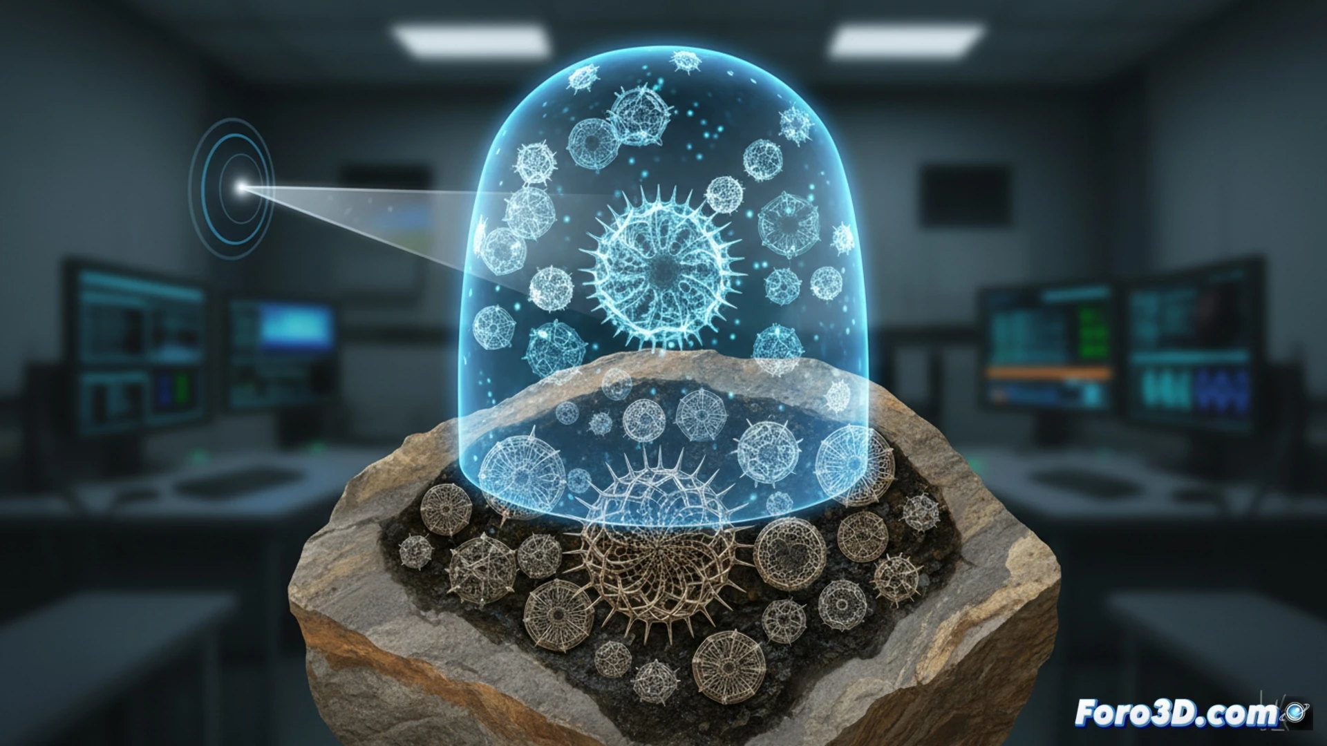
Synchrotron micro-tomography: a non-invasive window to the past 🔬
This technology was fundamental to the study. Instead of extracting and damaging the fragile fossils, the team used the synchrotron to obtain high-resolution 3D X-ray scans of the entire sample. The synchrotron light beam, extremely intense and focused, allowed for capturing the internal and external structure of each radiolarian with exquisite detail. Thus, three-dimensional digital models were generated that researchers could manipulate, measure, and study virtually without altering the valuable original, even identifying a species new to science.
3D visualization as a bridge for paleontology 🦴
This case exemplifies how scientific visualization techniques are revolutionizing disciplines like paleontology. The ability to observe the invisible without destroying, to create interactive digital replicas, transforms our understanding of the fossil record. These technologies not only unlock secrets of ancient biodiversity but also serve as a powerful tool for outreach, bringing extinct ecosystems to the screen with an unprecedented level of accuracy.
How can 3D scientific visualization techniques, such as high-resolution photogrammetry and volumetric rendering, be integrated to reconstruct and analyze ancestral microscopic ecosystems from fragmented fossil samples?
(PS: fluid physics for simulating the ocean is like the sea: unpredictable and you always run out of RAM)