A patient dies after the catastrophic failure of a bioprosthetic heart valve. Initially, a manufacturing defect is suspected. However, a forensic analysis with Micro-CT allows scanning the implant without removing it from the surrounding tissue. The resulting 3D model reveals the true cause: asymmetric calcification caused by an error in folding the biological material during surgery, not a device failure.
Technical workflow: from scanning to hemodynamic simulation 🛠️
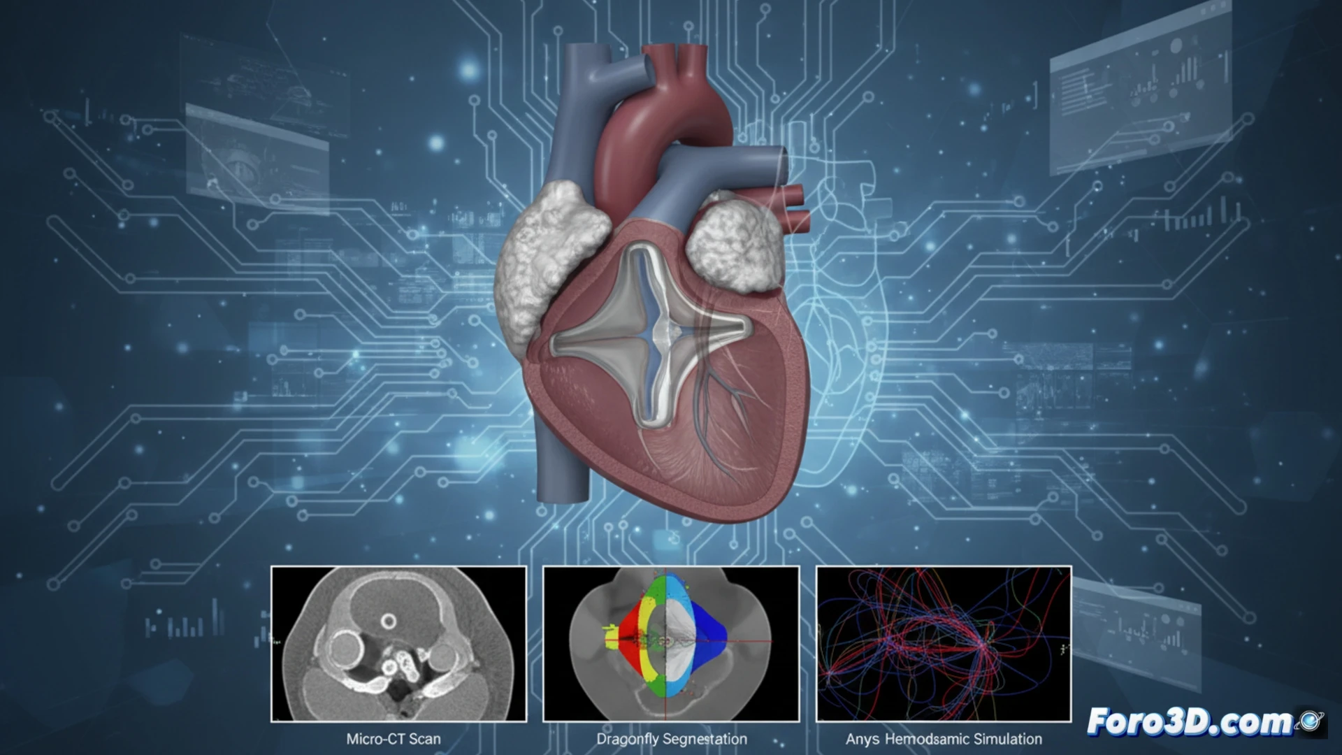
The process begins with scanning the tissue block on equipment such as Nikon CT or Zeiss Xradia, achieving micrometric resolution. Volume segmentation is performed in Dragonfly and 3D Slicer, separating the calcified tissue from the original valve structure. With the clean 3D model, it is exported to Ansys to run a hemodynamic simulation. This computational analysis demonstrates that the deformation from the surgical fold generated areas of high stress and turbulent flow, accelerating localized calcification. The workflow clearly differentiates an implantation error from a material defect.
Implications for forensic medicine and cardiac surgery 🔬
This case demonstrates that the combination of Micro-CT, advanced segmentation, and finite element simulation can turn a failed implant into a surgical learning tool. By identifying a folding error as the root cause, surgeons can review and standardize their implantation techniques. The methodology reinforces the value of 3D reverse engineering in forensic medicine, offering objective evidence to improve patient safety and reduce preventable failures in cardiac procedures.
How could the combination of micro-CT and 3D simulation transform the investigation of failures in bioprosthetic heart valves to prevent similar clinical tragedies?
(PS: If you 3D print a heart, make sure it beats... or at least that it doesn't cause copyright issues.)