The discovery of new variants of Osedax in the Southern Ocean reopens the debate on decomposition on the seafloor. These worms, known as zombie worms for their diet of whale bones, exhibit unique adaptations. For scientific visualization, representing their anatomy and life cycle in 3D is a fascinating challenge that allows understanding their role in nutrient redistribution in abyssal trenches.
Anatomical modeling and photogrammetry of specimens 🧬
To create an accurate representation, the first step is photogrammetry of real specimens preserved in ethanol. The base model of the adult worm shows a segmented body with a central trunk and red branchial plumes for respiration. The most complex part to model is the system of roots that penetrate the bone, where symbiotic bacteria reside. In 3D, the degradation of bone collagen can be simulated using displacement maps and translucent shaders for the tissue. Animating the life cycle, from the swimming larva to the sessile adult, requires advanced rigging to show the expansion of the roots and the release of sperm into the water, key processes for understanding their dispersal in the Antarctic ecosystem.
Ecological simulation and science communication 🌊
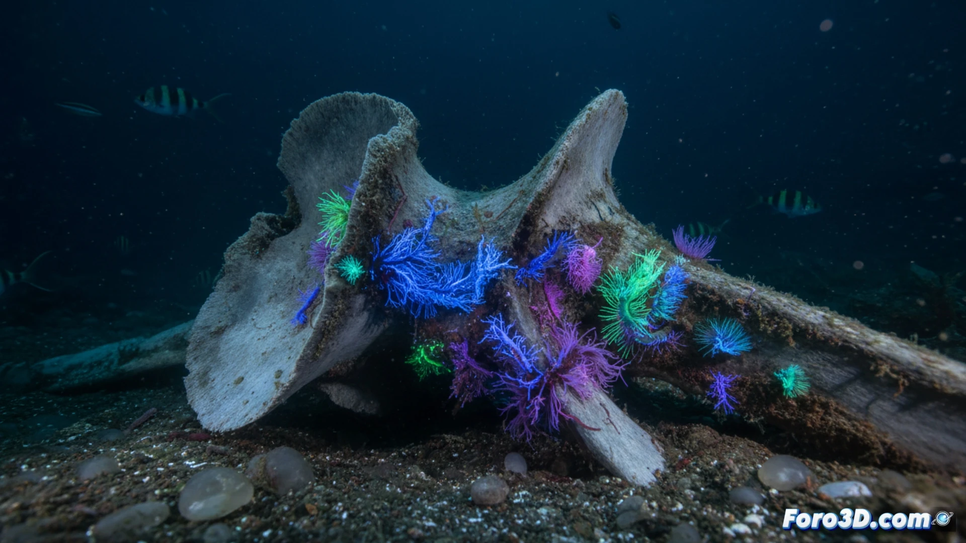
Beyond anatomy, 3D visualization allows simulating the whale fall ecosystem. Rendering the skeleton in a submarine canyon with deep-sea lighting helps contextualize the discovery. These tools are vital for science communication: by animating the colonization of the bone by multiple worms, researchers can show how these new variants compete for resources. Ultimately, 3D not only documents the species but reveals the hidden dynamics of life in the total darkness of the Southern Ocean.
As a 3D modeler, what technical and lighting considerations should I take into account to accurately simulate bioluminescence and the decomposition of a whale bone in the abyssal ocean when recreating Osedax?
(PS: fluid physics for simulating the ocean is like the sea: unpredictable and you always run out of RAM)